Radiology Case of the month: Right Adnexal Ectopic Gestation
Clinical History:
36 year old female with recent right ectopic gestation, Methotrexate given 1 week ago, presenting now with right lower quadrant pain.
Findings:
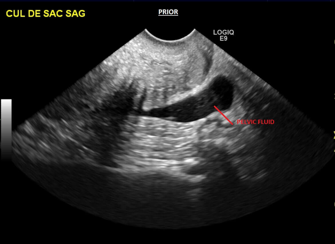
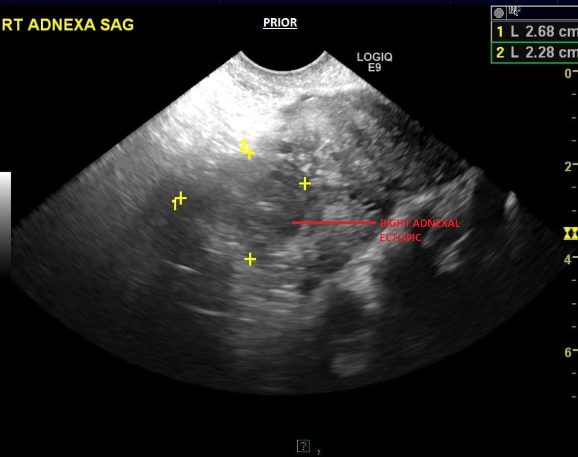
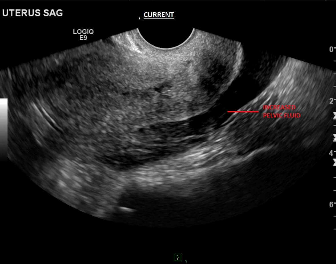
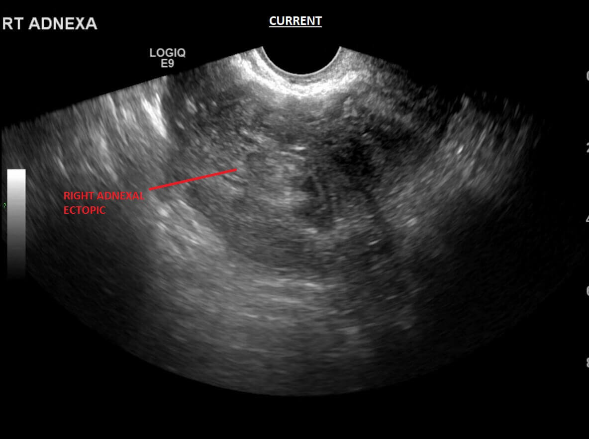
Pelvic ultrasound demonstrates a 2.8 x 2.3 x 2.4 cm complex lesion adjacent to the right ovary, similar to prior US examination. There is complex/hemorrhagic fluid in the pelvis, increased since the prior examination performed 7 days earlier.
Diagnosis:
Right adnexal ectopic gestation with interval increase in hemoperitoneum, concerning for tubal rupture.
Discussion:
Methotrexate is a folate antagonist that is given intramuscularly for treatment of ectopic pregnancy. A common regimen for methotrexate is a 50 mg/m2 dose that can be repeated on day 7 if the β-hCG value does not decline 15% between days 4 and 7. The requirements of methotrexate administration are hemodynamic stability, ultrasound findings consistent with an ectopic pregnancy, unruptured ectopic mass less than 3.5 cm in its greatest dimension, no fetal cardiac motion detected and β-subunit HCG level that does not exceed 5000 mIU/L.
On ultrasound, there may be an initial increase in the size of the tubal mass and increase in vascularity after methotrexate treatment. An adnexal mass may also be visible up to 3 months after treatment. The increase in tubal size and vascularity, in spite of the declining β-hCG level, represents a healing process and should not cause concern unless the patient is clinically unstable or has persistent symptoms.
Ultrasound after treatment with methotrexate is indicated in cases where rupture is suspected due to worsening abdominal pain or hemodynamic instability. Pain related to rupture should be differentiated from ‘separation pain’ caused by the separation of the pregnancy from the implanted site, which usually occurs 2-3 days after the injection. It is milder, of limited duration (lasting 24-48 h), and is not associated with signs of acute abdomen or hemodynamic instability. Ultrasound is also done when there is failure of β-hCG values to decline by at least 15% between days 4 and 7 or when increasing/plateauing β-hCG levels after the first week of treatment.
Take Home Points:
- Requirements of methotrexate administration are hemodynamic stability, ultrasound findings consistent with an ectopic pregnancy, unruptured ectopic mass less than 3.5 cm, no fetal cardiac motion and β-subunit HCG level not exceeding 5000 mIU/L.
- Ruptured ectopic is suspected after methotrexate treatment when there is worsening abdominal pain or hemodynamic instability.
References:
- Levine D. Ectopic pregnancy. Radiology 2007;245(2): 385–397.
- Bhatt, S., Ghazale, H., & Dogra, V. Sonographic evaluation of ectopic pregnancy. Radiologic Clinics of North America. 2007; 45:549-560.